Hysterosalpingography (HSG) or, in Turkish, “medicated uterus and tube film,” is a widely used method in gynecology to examine the structural and functional characteristics of the cervical canal, uterine cavity, and fallopian tubes.
A special fluid administered into the uterus allows these structures to be visualized on an X-ray film.
When is HSG performed?
In general, HSG is performed when there is suspicion of a blockage in the cervical canal, adhesions within the uterus, or a blockage in the fallopian tubes.
More specifically, the primary uses of HSG are as follows:
Determining whether the fallopian tubes are open during the medical evaluation of infertility
Evaluation of a possible obstruction in the cervical canal or adhesions in the uterus (Asherman syndrome) after a procedure performed on the cervix or uterus (such as abortion) that results in the cessation of menstrual bleeding
Determination of whether there is a congenital developmental defect in the uterus in cases of recurrent miscarriages
How is HSG performed?
HSG is generally performed during the first half of the menstrual cycle, after menstrual bleeding has completely stopped. This is to prevent problems caused by menstrual blood flowing from the uterus into the tubes and then into the abdominal cavity, and to avoid damaging a possible pregnancy in the uterus (the possibility of pregnancy should always be considered in women who are being evaluated for infertility).
A complete gynecological evaluation is performed before HSG. If signs of pelvic infection are detected during this evaluation, the procedure is postponed until the infection is treated. Performing an HSG while an infection is present may cause the infection to spread.
An HSG can be performed by a gynecologist or radiologist. The radiologist is responsible for providing the report.
While the patient is in the gynecological examination position, a device called a speculum is inserted to visualize the cervix. Once the cervix is stabilized, a cannula, which is a pipette-like device, is inserted into the cervical canal. A special liquid called a contrast medium is administered in several stages by applying pressure from a syringe attached to the cannula. This fluid makes the areas where it is located appear white on the X-ray film. While the fluid is being administered, X-ray films are taken at regular intervals, or a method called fluoroscopy is used to continuously monitor and record the passage of the fluid on a screen.
Although HSG is a method that uses X-rays, the radiation dose is not very high.
HSG is generally a procedure that causes minimal pain and is therefore not usually performed under general anesthesia.
How is HSG interpreted?
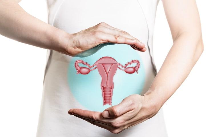
An HSG image is shown at the top of the page above. In this image, the uterus is seen in a triangular shape, with the vagina and cervical canal visible below the uterus. The capillary structures on either side of the uterus are the fallopian tubes, and beyond these structures, fluid can be seen spreading into the abdominal cavity. This image is an example of a completely normal uterus with both fallopian tubes open.
When no fluid reaches the uterus, this indicates a blockage in the cervical canal.
When “spots” are seen within the triangular structure of the uterine cavity, this indicates an adhesion within the uterus, and when the triangular structure of the uterine cavity is disrupted, this indicates a congenital uterine defect.
When the capillary structures (tubes) are not visible or when the distribution of fluid into the abdominal cavity is not observed, this indicates that the tubes are blocked.
What is done to resolve the issue identified during HSG?
Since fluid is administered under pressure during HSG, a slightly blocked cervical canal or fallopian tube may open under this pressure. Other than this, HSG has no therapeutic effect.
When Fallopian tube blockage is detected during HSG, IVF may be recommended directly, or laparoscopy may be performed to determine the cause of the adhesions in the tubes and treat them if possible.
If adhesions are detected in the uterus during HSG, there are different ways to resolve them, and hysteroscopy (examination of the uterus with a camera) is the most commonly used method, especially in advanced cases.
While it is possible to make a preliminary diagnosis of structural abnormalities in the uterus during HSG, definitive diagnosis typically requires hysteroscopy and laparoscopy. While it is possible to resolve the issue during these procedures, open surgery may be necessary for the treatment of certain congenital abnormalities.
We are here to provide the best service for your health. You can make an appointment by filling out the form below or calling our phone number.
Yeniköy Mah. Severcan Cad. Zeytindalı Önbahçe Evleri C Blok No:1 Bodrum
Monday - Friday: 09:00 - 18:00
Saturday: 09:00 - 13:00
Sunday: Closed